


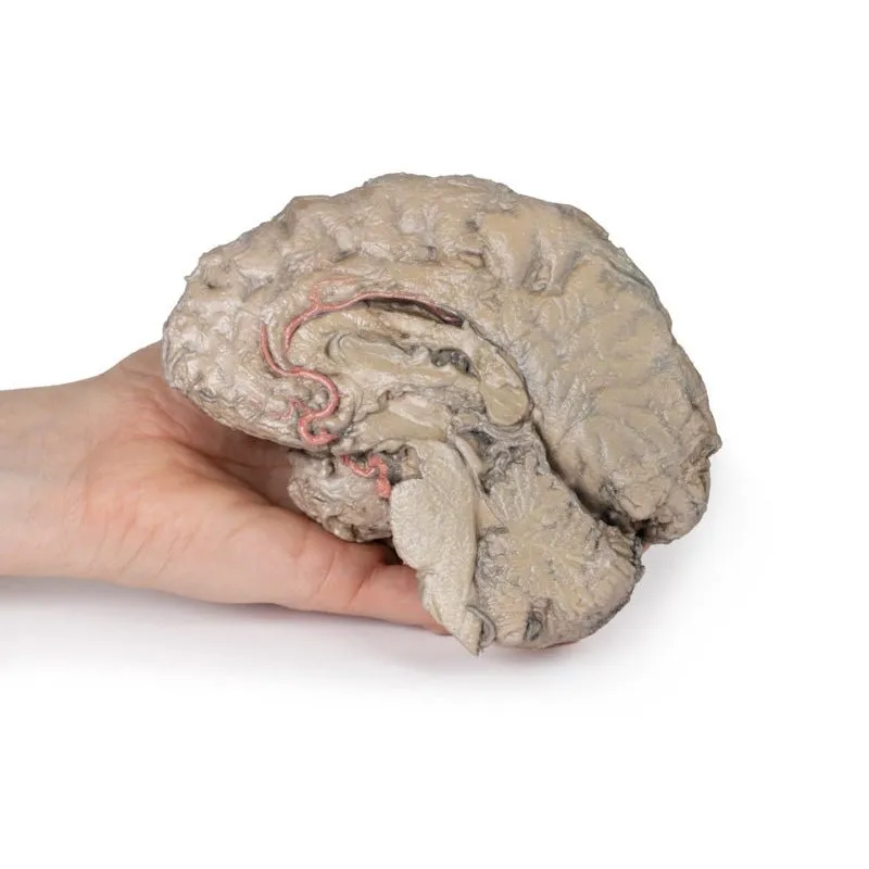
3D Printed Brain Hemisection
This 3D model is a midsagittal hemisection through a whole brain, preserving the right side anatomy and deep brain
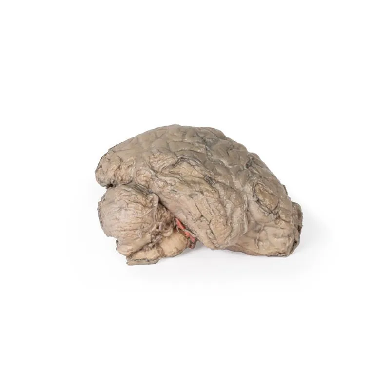
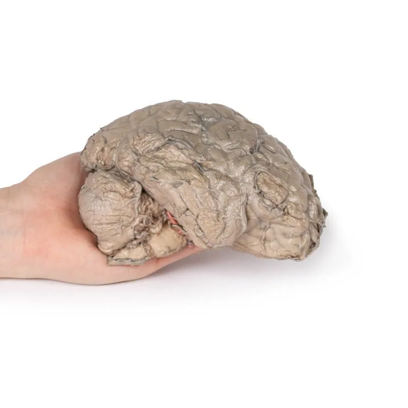
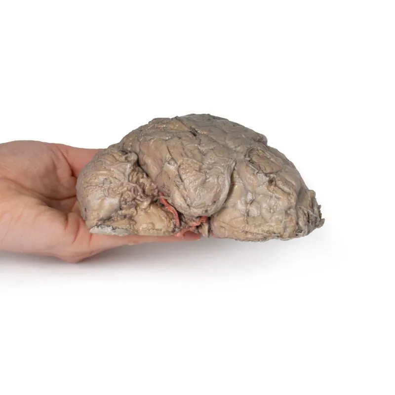
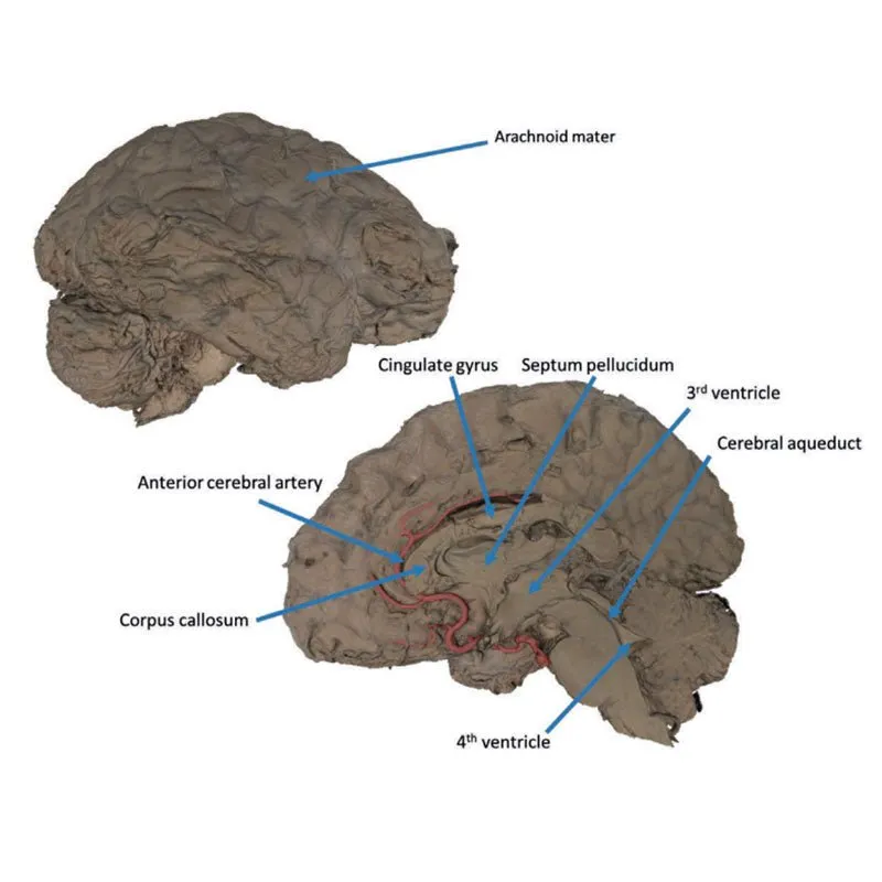
structures and spaces visible in the midline. In lateral view, the right cerebral and cerebellar hemispheres are
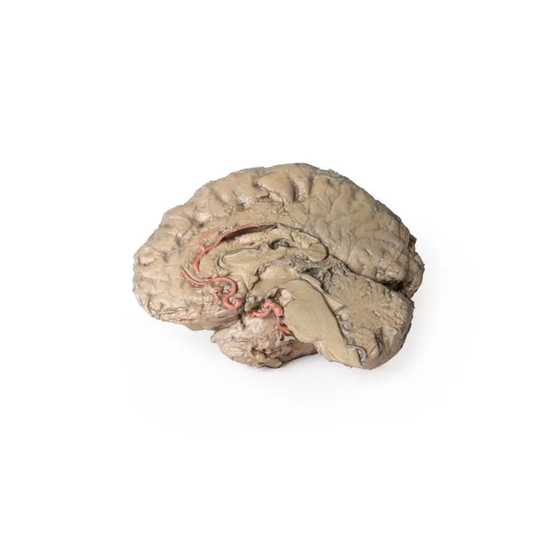
covered in the arachnoid mater. In the midline view, the brain regions from the cerebrum to the medulla oblongata
are preserved. Centrally, the third ventricle is opened, with an intact septum pellucidum superiorly positioned and
obscuring the lateral ventricles within the cerebral hemisphere. On the inferior margin of the third ventricle both
the right mamillary body and right optic tract can be observed, whereas posteriorly the cerebral aqueduct can be
observed extending across the midbrain between the tectum and tegmentum towards the fourth ventricle (between the
cerebellum and pons). The cerebellum is separated from the occipital lobe by a preserved portion of the tentorium
cerebelli, and in cross-section the cerebellar cortex helps form the prominent arbor vitae.
A series of arterial
branches have been false coloured to contrast their course across the preserved brain structures. In the midsagittal
view the anterior cerebral artery courses from around the corpus callosum to supply the cingulate gyrus and other
midline cortical regions. The base of the middle cerebral artery can be seen passing deep between the temporal and
frontal lobes, with the posterior communicating artery connecting it to a small remnant of the posterior cerebral
artery. Adjacent to the posterior cerebral is the superior cerebellar artery, extending laterally to pass between
the temporal lobe and the cerebellum before passing deep into the transverse fissure.
GTSimulators by Global Technologies
Erler Zimmer Authorized Dealer


